
I’ve been thinking about something a lot recently – proximal hamstring problems. (Seems like an exciting place to live – in my head, doesn’t it?) It all started with a number of patients that were seen in my medical clinic. In fact, over the course of a single week, I saw 7 patients with proximal hamstring tendon problems. During the week following – 4 more patients. Naturally, I began to wonder what in the world was going on here. First I questioned what it meant for all of these patients to have specifically sought care at my clinic for a literal pain in the butt. (Am I making more of that than I should?) In addition, I wondered what precipitating factors might have contributed to these cases.
While a few of the patients were athletes, most were not, and all experienced an insidious onset of symptoms. They described pain in the lower gluteal region, sometimes radiating down the posterior thigh. Pain was made worse by walking or running at a faster pace and by sitting for prolonged periods of time.
Ultrasound evaluations revealed a range pathology from mild hamstring tendinosis (chronic tendon degeneration) to frank tendon tears.
Let’s pause to review what we know about the hamstring muscles.
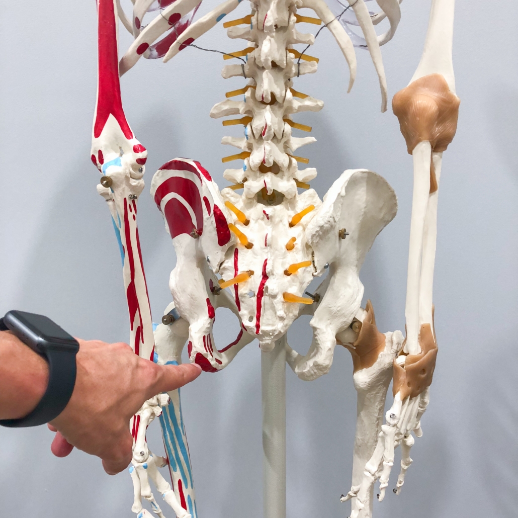
The hamstrings are a group of 3 muscles on the posterior thigh that collectively flex the knee and extend the hip (and are also technically involved in rotation of the leg). Can you name the 3 muscles? From medial to lateral, they are: semimembranosus, semitendinosus, and biceps femoris (which has a long and short head). The muscles originate from the ischial tuberosity (a.k.a. the “sit bone”) and insert past the knee joint on either the tibia or fibula bones.
Injuries to the proximal hamstring tendons do commonly occur during sports-related activities involving eccentric muscle contraction, forced hip hyperflexion, and full knee extension, but these injuries are often acute in nature. So what is to blame for the more chronic cases of proximal hamstring tendon pathology, especially in non-athletes?
When the problem of chronic proximal hamstring tendinopathy has been placed under a microscope (literally), studies have revealed pathologic changes in the tendon structure involving disruption of normal collagen architecture, increased abundance of extracellular matrix, and neovascularization (abnormal growth of new vessels). What causes these changes? Several studies have revealed many of the usual suspects of tissue degradation – inflammatory cytokines, matrix metalloproteinases (MMPs), various heat shock proteins, and a number of other malicious molecules. Let’s keep working backwards – But what causes the release of those molecules?
Want my answer?
Sitting.
I think that the many cases of chronic proximal hamstring tendinopathy that I see in my clinic can be attributed to prolonged sitting – something that we were never meant to do. (For more on the potential dangers of sitting, refer back to the May 2017 Muscle Meets Medicinearticle titled “Death by Chair?”)
Here’s the scoop:
When we sit, the soft tissues around the “sit bones” (the ischial tuberosities) are subject to significant increases in pressure. It is known that tissues are capable of sustaining pressure on the arterial side of around 30-32 mmHg for only a short duration. When pressures increase even slightly above this capillary filling pressure, it causes microcirculatory occlusion (impaired blood flow) and tissue ischemia, which results in the release of many of the aforementioned tissue-damaging molecules. In the same way that many hospitalized patients or otherwise immobilized persons are vulnerable to the development of skin ulcerations from prolonged periods of focal tissue pressure, so too are other soft tissues adversely affected by capillary compression.
(Also notable: There are likely some genetic risk factors that predispose certain individuals to developing tendinopathies when other given conditions are present. For more on this, you can search polymorphisms of the COL5A1 gene.
Even more: It was demonstrated in a study by Ruzzini et al. that tendon stem cells isolated from older hamstring tendons had less potential for tissue healing, thus suggesting a likely biological basis for age-related differences in tendon integrity.)
Most of the patients seen in my clinic for proximal hamstring tendinopathy have been treated with a regenerative procedure (such as platelet rich plasma or amniotic allograft administration) in order to encourage tendon healing. This is done in combination with biomechanical evaluation and recommendation of specific therapeutic exercises. (And ultimately, a lecture on the dangers of prolonged sitting…)

